Sputum smear microscopy in the detection of tuberculosis
Tuberculosis has remained a global health burden, causing morbidity and mortality in millions of people worldwide.
The rapid detection of infection is critical in the management and control of disease spread. Towards case detection of tuberculosis, sputum smear microscopy continues to remain highly relevant.
In fact according to the World Health Organisation guidelines on the management of tuberculosis, a vital aspect of patient investigation in suspected cases of tuberculosis is the examination of the sputum samples under a microscope.
Also, the International Standards for Tuberculosis states that every patient whether a child, an adolescent, or an adult, who is able to produce sputum and is a tuberculosis suspect should produce two or ideally, three samples for microscopic examination.
Even though other means of tuberculosis detection exist, due to its simplicity, rapidity, and relatively low cost, sputum microscopy continues to be indispensable in the diagnosis of tuberculosis. Added to this is its high specificity for mycobacterium tuberculosis, by being able to differentiate between it and the non-tuberculous mycobacterium.
Microscopy of acid-fast bacilli – stained sputum is positive in more than 75% of adults and less so for paediatric cases, and while this may be less sensitive than other methods like sputum culture, some procedural measures can be taken to further enhance the sensitivity of sputum microscopy in tuberculosis case detection.
Some of these measures include:
- Careful sample collection
- Good smear and staining technique
- Centrifugation for higher bacilli concentration
- Bleach processing and sedimentation for a clearer microscopy field
- The use of different concentrations of reagents, etc.
Not only is sputum microscopy highly specific in areas of high tuberculosis prevalence, but it is also able to identify the most infectious patients, and is applicable across a wide range of populations.
It is not surprising therefore, that it has remained a vital aspect of the global strategy for the control of tuberculosis.
From a realistic point of view, it may be a reasonable length of time before newer diagnostic methods like the gene Xpert molecular rapid diagnostic test would be readily available as a replacement for microscopy, especially in areas where there is a high prevalence of HIV and tuberculosis co-infections, or multidrug-resistant tuberculosis.
The efficient diagnosis of tuberculosis ideally would require a test that has the following features:
- Widely available even in limited resources situations
- Has no need for electricity or refrigeration
- Has no need for clean water access
- Is user-friendly with little or no training required
- Quick turnaround of results
- High sensitivity and specificity
- High negative and positive predictive values
- Remain relevant over a long period of time.
Currently, while no diagnostic test presently fulfills all the criteria, the sputum smear microscopy comes closest to doing so and therefore, until such an ideal diagnostic test is developed, remains the commonest, most practical, and cost-effective diagnostic test for tuberculosis globally.
The continued relevance of sputum smear microscopy notwithstanding, there are certain limitations associated with its use such as the time-consuming nature of having to manually read slides, and user influenced errors affecting the quality of results.
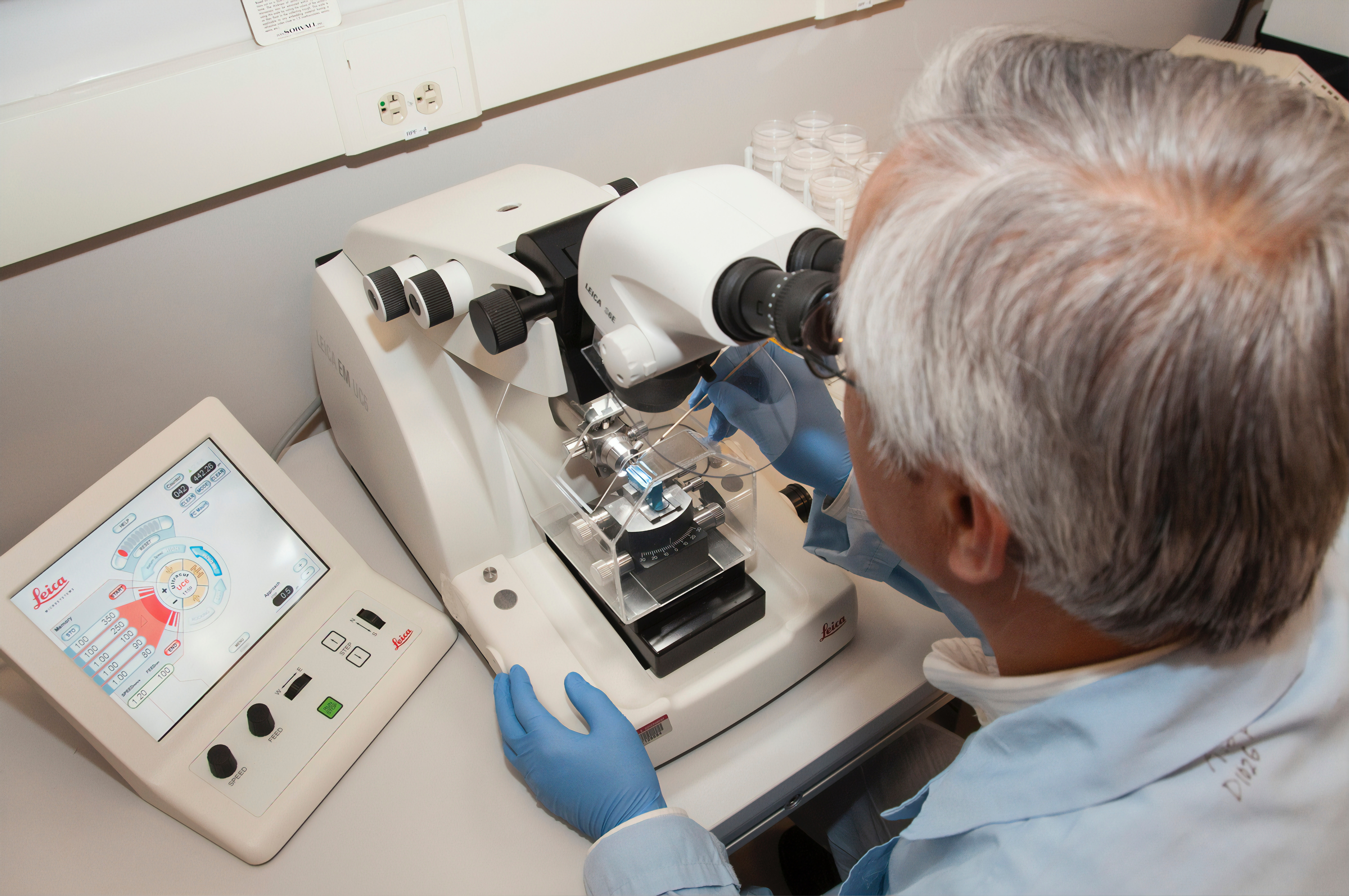
These limitations are greatly minimised or even removed with the introduction of the microscopy automation platform, which is the use of high-resolution and sensitive microscopes that are able to scan and analyse several hundred slides in a single batch, effectively leading to a significant improvement in sputum smear microscopy workflows, reduction of labour costs, and increased cost effectiveness.
Not only is the automation platform microscopy useful in tuberculosis diagnosis, but also in automated gram stain reading and direct multiplex imaging used in the direct detection of pneumonia-causing micro-organisms.
Automated smear microscopy achieves a higher sensitivity and specificity compared with the conventional microscopy and culture results and therefore, has a great potential in filling the existing urgent need for a rapid, large-scale screening tool for tuberculosis.


